

Guidelines in Practice Webinar Series
Please join us for our new Guidelines In Practice webinar series!
November 3, 2025 • JASE
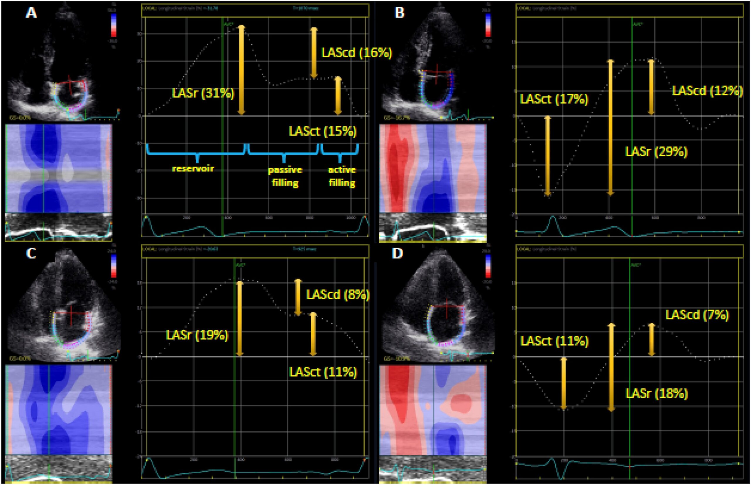
Clinical Applications of Strain Echocardiography
Clinical Applications of Strain Echocardiography: A Clinical Consensus Statement From ASE Developed in Collaboration With the EACVI
November 2, 2023 • JASE
COVID-19 Statement Update
ASE COVID-19 Statement Update: Lessons Learned and Preparation for Future Pandemics
January 27, 2021 • JASE
Adapting Pediatric, Fetal and CHD Services COVID-19
ASE Statement on Adapting Pediatric, Fetal, and Congenital Heart Disease Echocardiographic Services to the Evolving COVID-19 Pandemic
May 19, 2020 • JASE
Reintroduction of Echo During COVID-19
ASE Statement on the Reintroduction of Echocardiography Services During the COVID-19 Pandemic
April 7, 2020 • JASE
ASE Statement on COVID-19
ASE Statement on Protection of Patients and Echocardiography Service Providers During the 2019 Novel Coronavirus Outbreak: Endorsed by the American College of Cardiology
November 1, 2005 • JASE
Minimum Standards for the Cardiac Sonographer
American Society of Echocardiography Minimum Standards for the Cardiac Sonographer: A Position Paper
October 1, 2004 • JASE
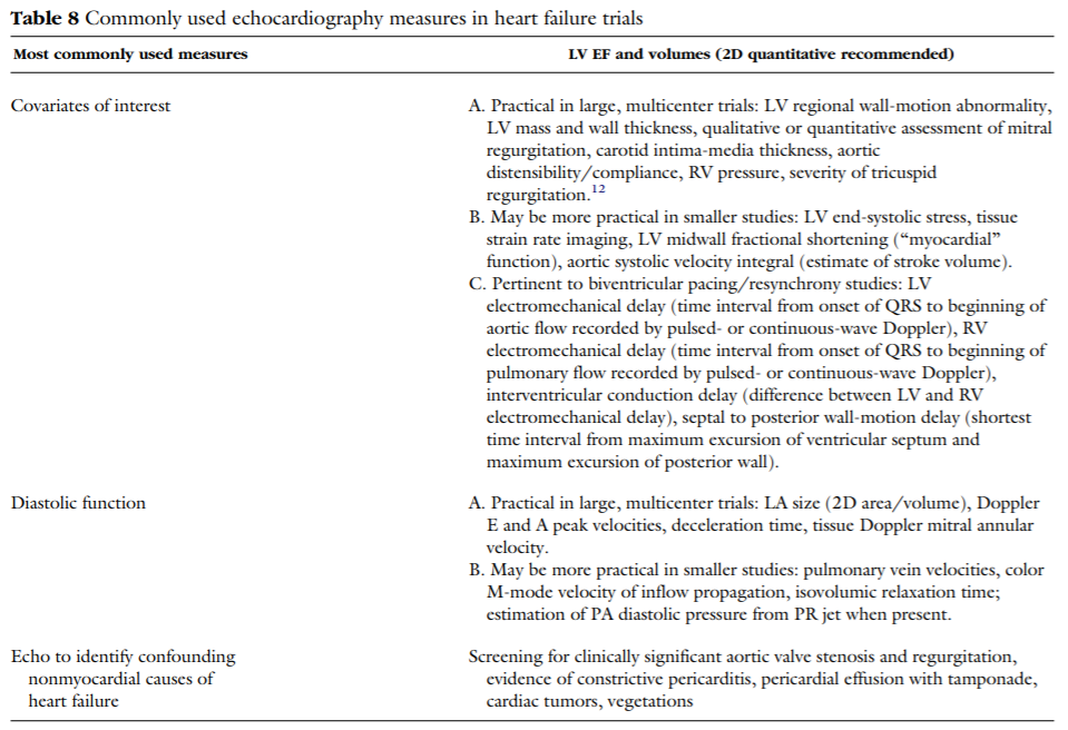
Echo in Clinical Trials
American Society of Echocardiography Recommendations for Use of Echocardiography in Clinical Trials
Read the 2026 ACC/AHA/ASE/HRS/STS Expert Consensus Systems of Care Document
Guidelines in Practice Webinar Series

Guideline Webinar
Guidelines In Practice Webinar Series: Ultrasound Artifacts
Please join us for the 3rd webinar in...
July 14, 2026 • 06:00 pm America/New_York