
 The latest issue of CASE is now available with intriguing reports, including “Aortic Valve Myxoma in a Young Child.” CASE Editor-in-Chief Vincent Sorrell, MD, FASE, remarked, “Masses in the heart are relatively common and the learned echocardiographer is aware of the many characteristic features to help create a differential diagnosis, recognizing that, in the end, a final diagnosis requires tissue. Most importantly, categorization as a benign (often normal variant or embryologic remnant) or a pathologic mass is the first step. Once the visualized mass is established as pathologic, then the clinical setting (patient characteristics, signs, and symptoms) is essential to drive further testing or treatment. These authors present a fascinating case of a five-year-old, asymptomatic girl with a murmur found to have a very unusual, large (> 1.0 cm) mass attached to the ventricular surface of the aortic valve non-coronary cusp. The authors provide excellent images and walk the reader through their consideration for potential etiologies (including Carney complex) and role for advanced imaging. Given the size of the mass and partial obstruction (transvalvular gradient nearly 25mmHg) surgery was performed and confirmed that the mass was a myxoma. This is another nice example of how CASE reports can help expand our diagnostic considerations.”
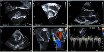
Continuing in the Valvular Heart Disease category, Oursbourn et al. report their uncommon findings of multi-valvular fulminant infective endocarditis in a healthy pediatric patient after an initial misdiagnosis of acute rheumatic fever. Limaye et al. provide exceptional images of the unusual Doppler pattern in their patient who was found to have a complex fistula involving the sinus of Valsalva. Cardiac Tumors and Pseudotumors supplies readers with two cases: Xuqian et al. share excellent contrast perfusion images in their report of a teenager with dyspnea and dizziness who was found to have a left atrial myxoma, while Khaleel et al. present two cases of incidentally found left atrial masses during LAAO device placement TEE imaging. Rounding out this issue is Just Another Day in the Echo Lab featuring a report on the use of STE bullseye pattern in a patient with confirmed acute pulmonary embolism and the associated echo finding of RV systolic dysfunction with focal apical sparing. Finally, Dr. Sorrell’s editorial shares behind-the-scenes commentary and insights from each of the published CASE reports.
SUBMIT your case report to us! Whether it will be your first time submitting a case or your 50th, we are here to make it a great experience. Email us with questions or submit your report today!
Related News

Beyond the Blockage: Coronary Artery Disease with a Twist
February 18, 2026
New Issue of CASE is Brrrrr-illiant
January 21, 2026
Gobble Up a New Issue of November CASE
November 18, 2025