The latest focus issue of CASE is now available with intriguing reports, including “The Hidden Constriction: Middle Aortic Syndrome Presenting as Acute Systolic Heart
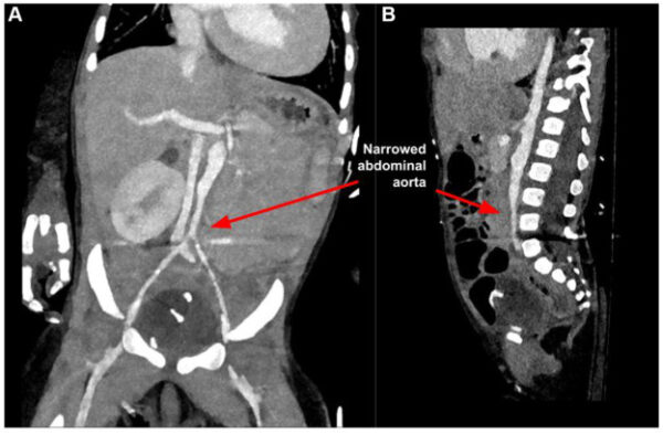
Failure in a 7-Month-Old Infant,” by Zhang Et Al. CASE Editor-In-Chief, Vincent Sorrell, MD, FASE, remarked, “In another striking example of the resilience of our pediatric population that make up an increasing proportion of manuscripts published in CASE, these authors described their 7-month-old infant with middle aortic syndrome (MAS). As an adult cardiologist, this was another entirely new syndrome for me, and I learned a lot. This baby presented to the emergency department with a striking blood pressure of 159/128mm Hg. Echocardiography demonstrated severely reduced LV systolic function and moderate functional MR, but the visualized portions of the aorta were normal without coarctation. However, the computed tomography scan demonstrated MAS with a 2.1cm length of abdominal aorta narrowed to approximately 3-4mm. This child has been medically managed, and the authors include an excellent discussion that is sure to pique your interests. For their report, they also included the follow-up TTE with recovered LV function”.
This focus issue continues to take readers on a tour through the power of multimodality imaging, demonstrating how each modality contributes a unique piece of to the diagnostic puzzle. Kumar Sahoo Et Al. showcases a true imaging relay race, with echocardiography identifying severe coarctation, CCT guiding intervention and invasive angiography uncovering an acute aortic dissection. Solem Et Al. illustrates how echocardiography and CMR can work hand in hand to distinguish a tumor from a thrombus, while Hasegawa Et Al. elegantly demonstrated how CCT can further define the origin and venous extension of a left atrial mass first detected by echocardiography. Brinchiro Et Al. continues the theme of complementary imaging, highlighting the value of CT in investigating abnormal pulmonary venous flow identified on echocardiography. Huang Et Al. emphasizes the indispensable role of echocardiography in diagnosing complex congenital heart disease, while illustrating how CCT can help chart the course for surgical intervention. Rounding out the issue, Durrwachter Et Al. proves that the benefits of multimodality imaging extends beyond human medicine, combining echocardiography and CT to diagnose a rare coronary anomaly in a dog. Together, these cases highlight how today’s imaging toolbox allow clinicians to see the full picture.
Readers can look forward to Dr. Sorrell’s editorial, “Sound Waves, Rays, and Magnets”.
Want to challenge your imaging and diagnosis skills, visit the CASE Homepage to see the latest Unlock the CASE image quiz as well as a brand-new Sonographer Sound-Off.
Share your next great case with CASE! Whether you’re preparing your very first submission or adding to a long list of published reports, we are here to make it a great experience. Email us with questions or submit your report today!