Recognize ASE Members Going the Extra Mile

Gemstones or “gems” are highly valuable, and similar to their namesake, ASE GEMs recognize valued members who Go the Extra Mile.

Join ASE in celebrating members who are making the world a better place by submitting a GEM nomination. Nominate a colleague for their dedication to the field of cardiovascular ultrasound, outstanding mentorship to students or fellows, commitment to quality patient care, or congratulate them on a major accomplishment or milestone service anniversary.

The deadline for submission is the 25th of each month to allow time for recognition the following month. View past ASE GEM recipients here.

Register Now for the 24th Annual ASCeXAM/ReASCE Review Course

Content is available now for the 24th Annual ASCeXAM/ReASCE Review Course. This course is specifically designed to help you gain the knowledge you need to pass the ASCeXAM® and ReASCE® exams, but even if you are not taking the exam this year, this course offers comprehensive cardiovascular ultrasound education. In addition to covering all aspects of cardiovascular ultrasound essential to these examinations, your registration also includes a virtual live component coming up on Saturday, July 8. The expert faculty will host two, two-hour Q & A sessions from 10:00 AM – 12:00 PM ET and 12:30 – 2:30 PM ET. Register today, start studying, and earn 28.75 AMA PRA Category 1 Credits™ (pending approval).

Once In a Blue Moon: A Rare CASE for June

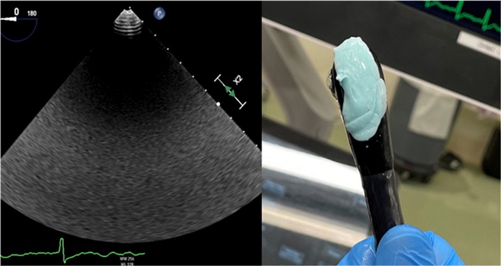
The latest issue of CASE, ASE’s open access case reports journal, is now available with some fascinating reports, including “A Sticky Situation: The Unfortunate Consequence of Chewing Gum” by Christopher Lee, MD et al. CASE Editor-in-Chief, Vincent L. Sorrell, MD, FASE, remarked, “This issue includes a rarity! Since assuming the role as EIC, I can honestly say that this has never occurred in the many hundreds of manuscripts I have reviewed. In this issue, there is one of the most clinically meaningful CASE reports we’ve published, and it did not include any high-quality images. In fact, there were no interpretable images whatsoever. And that is the point of their report. Lee et al. should be congratulated in their careful approach to a patient with grossly impaired acoustic transmission during a TEE procedure.” Read the full article in CASE to learn more!

Additionally, there are two reports in the Multimodality Imaging category and three in the Interventional Echocardiography category, including a case on the serial evaluation of the oldest living patient with asymptomatic severe apical variant hypertrophic cardiomyopathy. A diverse range of reports fall into the Congenital Heart Disease category, including one from a 38-year-old with an untreated Gerbode defect who underwent coarctation stenting. In his editorial, Dr. Sorrell parallels his travels westward to the daily explorations of echocardiographers and the images that tell a story if one looks close enough.

Looking for a journal to submit your case report to? We want to hear from you! Email us with questions or submit your report today. Be sure to check out the latest Sonographer Sound-Off and Unlock the CASE features on the CASE Homepage to view more captivating work from your colleagues.

ASE 2023 Research Spotlight 4

FOR IMMEDIATE RELEASE

Contact: Angie Porter
919-297-7152
APorter@ASEcho.org

Advances in Echocardiography Lead to Promising Research for Placing Pacemaker Devices

(DURHAM, NC, June 23, 2023)—Research that will be presented at the American Society of Echocardiography’s (ASE) 34th Annual Scientific Sessions, June 23-26, 2023, in National Harbor, Md., will share how advances in echocardiography resulted in more accurately placed pacemaker devices.

Some pacemakers use flexible, insulated wires—also known as leads—to deliver electrical pulses that help hearts beat at a normal rate and rhythm. The current standard practice for placing pacemaker leads is fluoroscopic/X-ray guidance. However, a new research study conducted by Children’s Hospital Colorado in Aurora, Colo., found that three-dimensional (3D) echocardiography resulted in more accurate pacemaker lead placement when compared to X-ray technology.

The study’s lead author Dr. Dale Burkett, who is an Assistant Professor in Pediatrics-Cardiology at Children’s Hospital Colorado, explained that when pacemaker leads are placed in the ideal location, they are able to better mimic the heart’s normal electrical conduction and reduce the long-term risk of heart dysfunction. Additionally, 3D echocardiography lowers both the patient’s and the cardiology staff’s exposure to radiation during the procedure.

“By using 3D echocardiography guidance, we are able to better visualize pacemaker leads as they move through the heart and guide them to where they are intended to go,” says Dr. Burkett. “Our work demonstrates that with advances in echocardiography, we can provide higher quality care for our patients who need permanent pacemakers for heart rhythm management.”

Highlighting continuing innovations in the cardiovascular ultrasound field, nearly 400 abstract poster presentations will be featured during ASE 2023. This study, titled 3D Echocardiography Guidance for Pacemaker Lead Placement Improves Accuracy of Lead Placement and Reduces QRS Duration Compared to Fluoroscopic Guidance, will be presented Sunday, June 25, 4 p.m. to 4:45 p.m. ET at the Gaylord National Harbor Resort and Convention Center. Learn more about ASE 2023 and download the conference’s final event program at ASEScientificSessions.org.

About American Society of Echocardiography
The American Society of Echocardiography (ASE) is the Society for Cardiovascular Ultrasound Professionals™. ASE is the largest global organization for cardiovascular ultrasound imaging serving physicians, sonographers, nurses, veterinarians, and scientists and as such is the leader and advocate, setting practice standards and guidelines for the field. The Society is committed to advancing cardiovascular ultrasound to improve lives. For more information, visit ASE’s website ASEcho.org or social media pages on Facebook, Twitter, LinkedIn, or Instagram.

###

ASE 2023 Research Spotlight 3

FOR IMMEDIATE RELEASE

Contact: Angie Porter
919-297-7152
APorter@ASEcho.org

Artificial Intelligence in Cardiovascular Imaging Research Highlighted at ASE 2023

(DURHAM, NC, June 23, 2023)—Artificial intelligence (AI) is transforming the field of cardiovascular ultrasound imaging, and research studies are examining the ways that AI can most benefit patients and medical practitioners.

AI-related research is a prominent topic at the American Society of Echocardiography’s (ASE) 34th Annual Scientific Sessions, June 23-26, 2023, in National Harbor, Md. This year’s meeting is appropriately named Foundations and the Future of Cardiovascular Ultrasound and will focus on education, science and innovation.

Three AI research studies that will be presented during the meeting highlight different AI tools including AI-powered chat interactions, identifying patients with heart failure with preserved ejection fraction (HFpEF) and identifying life-threating valve disease. The findings from each of these studies will be presented Sunday, June 25, 2 p.m. to 2:45 p.m. at the Gaylord National Harbor Resort and Convention Center. Learn more about ASE 2023 and download the conference’s final event program at ASEScientificSessions.org.

Using AI-Powered Chat Interactions with Patients

The rise of online chat-based AI models has opened up new possibilities in the field of medicine. A new research study titled Enhancing Patient Comprehension of Echocardiography Reports through Artificial Intelligence-Powered Chat Interactions examines how chat-based AI technology could potentially improve the healthcare experience for cardiovascular ultrasound patients.

Echocardiography reports are crucial for diagnosing and monitoring heart conditions, but they are oftentimes filled with complex medical terminology and numerous abbreviations that can be difficult for patients to understand. Providers use these reports to explain medical findings to patients, and patients can use the reports to actively engage in their healthcare, participate in insightful discussions with their doctors and obtain a better comprehension of treatment strategies.

“Our research is fundamentally about patient education and bridging the gap between medical complexity and patient understanding,” says lead author Dr. Joseph Kassab, a cardiovascular imaging research fellow at Cleveland Clinic in Ohio. “By leveraging AI-powered chat interactions, it is our hope that in the future, we might be able to provide patients with a medically validated tool to explore and better understand their echocardiography reports on their own time. Doctors continue to provide their irreplaceable expertise, while the AI models serve as a supportive educational tool, making information more accessible to patients.”

AI Can Detect HFpEF with Greater Ease and Precision

Heart failure with preserved ejection fraction (HFpEF) is a major cause of disease and death worldwide, and diagnosis can be challenging. Various algorithms that include clinical risk factors and echocardiography measurements have been developed for diagnosis but are indeterminate in many patients. A research study titled Automated Echocardiographic Detection of Heart Failure with Preserved Ejection Fraction using Artificial Intelligence applies AI to analyze a single, routinely acquired echocardiographic video clip in an effort to identify patients with HFpEF, a group with increased morbidity and mortality.

“Our novel AI model demonstrated excellent discrimination of patients with HFpEF versus without, more often than with clinical scores, and it was able to stratify patients according to mortality risk,” said the study’s senior author Dr. Patricia A. Pellikka, chair, Division of Cardiovascular Ultrasound at Mayo Clinic in Rochester, Minn. “Further testing in varied populations and in different echocardiography labs is needed to refine the model, but it could potentially be used for screening and importantly, expedite diagnosis and treatment for a large group of patients.”

The deep learning-based AI algorithm was developed as a result of institution and industry collaboration. Mayo Clinic and Ultromics, an AI echocardiography company spun out of the University of Oxford, joined forces to develop the algorithm, which was cleared by the Food and Drug Administration (FDA) in 2022 after being awarded breakthrough device status. The research was supported by a grant from the ASE Foundation.

AI Technology Supports Echocardiography in Determining Aortic Stenosis Severity

Aortic Stenosis (AS) is a common form of valvular heart disease, and it is estimated to be present in over 12% of the population aged 75 years and above. Transthoracic echocardiography is the first line of cardiovascular imaging in determining the severity of AS and requires expert sonographic and interpretation capabilities to yield accurate results. AI technology has emerged as a useful tool to address these limitations but has not yet been applied in a fully hands off manner to evaluate AS.

A new research study titled Fully Automated Artificial Intelligence Assessment of Aortic Stenosis by Echocardiography tested an AI tool to measure echocardiographic markers of AS severity without human input. The AI-derived measurements were highly accurate when compared against expert cardiologist assessment.

“AI technology can allow for near instantaneous and hands-free identification of a life-threatening valvular heart condition by providers trained only in basic ultrasound,” said lead author Dr. Hema Krishna, a cardiologist at the University of Illinois at Chicago. “This machine learning platform can potentially extend diagnostic capability to patients in rural, community, or emergency room settings without immediate access to cardiologists.”

Dr. Krishna explained that rapid identification of hemodynamically significant AS can reduce the time to receive life-saving valve replacement therapy and also prevent unnecessary morbidity and mortality.

About American Society of Echocardiography
The American Society of Echocardiography (ASE) is the Society for Cardiovascular Ultrasound Professionals™. ASE is the largest global organization for cardiovascular ultrasound imaging serving physicians, sonographers, nurses, veterinarians, and scientists and as such is the leader and advocate, setting practice standards and guidelines for the field. The Society is committed to advancing cardiovascular ultrasound to improve lives. For more information, visit ASE’s website ASEcho.org or social media pages on Facebook, Twitter, LinkedIn, or Instagram.

###

ASE 2023 Research Spotlight 2

FOR IMMEDIATE RELEASE

Contact: Angie Porter
919-297-7152
APorter@ASEcho.org

New Research Study Uses Echocardiography to Compare Global Longitudinal Strain Values Among College Athletes Diagnosed with a COVID-19 Infection

(DURHAM, NC, June 23, 2023)—COVID-19 may no longer be a global health emergency, but the effects of the disease, which is caused by the SARS-CoV-2 virus, are still being felt and studied. Research that will be presented at the American Society of Echocardiography’s (ASE) 34th Annual Scientific Sessions, June 23-26, 2023, in National Harbor, Md., will share how echocardiography was used to analyze left ventricular global longitudinal strain (GLS) in college athletes who experienced mild-to-moderate SARS-CoV-2 infection.

In the study, nearly 200 student athletes from the University of Wisconsin-Madison, who tested positive for COVID-19 from September-March 2020, underwent a comprehensive echocardiogram to collect various data, including GLS values. GLS values can vary with age and sex, but they can be used to help predict future cardiovascular events and detect structural heart diseases.

The study found that GLS was normal in most college athletes when compared to reported normal values. However, GLS values in male football players were lower than previously reported normal values. These echocardiographic findings were supported by cardiac MRIs.

“Collecting and comparing GLS enables medical professionals to better monitor athletes for heart abnormalities associated with viral illnesses, such as COVID-19,” says the study’s lead author Dr. Parag A. Tipnis, an associate clinical professor in the Division of Cardiovascular Medicine at the University of Wisconsin-Madison. “Echocardiography remains a powerful tool in the diagnosis of cardiac disease and can be complementary to other modalities, such as cardiac MRIs, to help determine when athletes or patients can return to normal activities after an illness.”

Dr. Tipnis described the research findings as hypothesis generating, and stressed the importance of further analyzing GLS values in larger patient populations.

“My research team and I believe that future studies of GLS patterns in college athletes will be important in helping detect heart issues and minimizing the risk of sudden cardiac death,” he continued.

Highlighting continuing innovations in the cardiovascular ultrasound field, nearly 400 abstract poster presentations will be featured during ASE 2023. This study, titled Comparison of Global Longitudinal Strain Among College Athletes Diagnosed with Mild to Moderate SARS-CoV-2 Infection, will be presented Saturday, June 24, 7 a.m. to 7:50 a.m. ET at the Gaylord National Harbor Resort and Convention Center. Learn more about ASE 2023 and download the conference’s final event program at ASEScientificSessions.org.

About American Society of Echocardiography
The American Society of Echocardiography (ASE) is the Society for Cardiovascular Ultrasound Professionals™. ASE is the largest global organization for cardiovascular ultrasound imaging serving physicians, sonographers, nurses, veterinarians, and scientists and as such is the leader and advocate, setting practice standards and guidelines for the field. The Society is committed to advancing cardiovascular ultrasound to improve lives. For more information, visit ASE’s website ASEcho.org or social media pages on Facebook, Twitter, LinkedIn, or Instagram.

###

ASE 2023 Research Spotlight 1

FOR IMMEDIATE RELEASE

Contact: Angie Porter
919-297-7152
APorter@ASEcho.org

Large Study Finds Artificial Intelligence Can Assist Novice Nurses, Medical Residents in Acquiring Diagnostic-Quality Echocardiography Images

(DURHAM, NC, June 23, 2023)—Artificial intelligence (AI) is changing the way echocardiograms are acquired and interpreted. A new study, that will be presented at the American Society of Echocardiography’s (ASE) 34th Annual Scientific Sessions, June 23-26, 2023, in National Harbor, Md., will share how AI-guided software can benefit both the field of cardiovascular ultrasound and ultimately its patients.

The multi-center study compared the quality of echocardiography images acquired by nurses and medical residents. After just 12 hours of training with the AI-guided software, these novice users were able to acquire echocardiography images in 10 standard views with rates of diagnostic quality approaching that of expert sonographers.

The AI-guided software was developed by UltraSight using intuitive visual cues from more than 10,000 echocardiograms and was embedded into a Philips Lumify handheld device. The accuracy of the AI software was then evaluated by five expert readers who were blinded to the imagers’ identities and evaluated for sufficient quality for diagnostic interpretation.

“We found that the AI-guided software allowed novices to acquire images suitable for diagnostic interpretation by an expert reader in the majority of patients,” says one of the study’s lead authors, Dr. Roberto M. Lang, an internationally renowned cardiologist and specialist in echocardiography at the University of Chicago Medicine. “These promising findings confirm that AI-guided software could be potentially useful in teaching novice medical professionals and assessing cardiac function in settings where services of trained echocardiography laboratories are not available.”

Nearly 400 abstract poster presentations highlighting continuing innovations in the cardiovascular ultrasound field will be featured during ASE 2023. This study, titled Real-Time Artificial Intelligence Based Guidance of Echocardiographic Imaging: Does Novice Profile Affect Image Quality and Suitability for Diagnostic Interpretation? will be presented by University of Chicago Non-Invasive Cardiovascular Imaging Fellow Dr. Juan Ignacio Cotella on Sunday, June 25, from 3 p.m. to 3:45 p.m. ET at the Gaylord National Harbor Resort and Convention Center. Learn more about ASE 2023 and download the conference’s final event program at ASEScientificSessions.org.

About American Society of Echocardiography
The American Society of Echocardiography (ASE) is the Society for Cardiovascular Ultrasound Professionals™. ASE is the largest global organization for cardiovascular ultrasound imaging serving physicians, sonographers, nurses, veterinarians, and scientists and as such is the leader and advocate, setting practice standards and guidelines for the field. The Society is committed to advancing cardiovascular ultrasound to improve lives. For more information, visit ASE’s website ASEcho.org or social media pages on Facebook, Twitter, LinkedIn, or Instagram.

###

Congratulations to ASE’s Newest FASE

This month, ASE welcomed 15 distinguished Fellows of the American Society of Echocardiography (FASE) recipients! These members exemplify excellence in the field of cardiovascular ultrasound through education, research, leadership, and volunteerism. They are ambassadors for ASE and recognized experts in their field. Join us in congratulating the newest FASE recipients:

  • Cecilia A. Albaro, MD, FASE
  • Mounir F. Banoub, MD, FASE
  • Sridhar Banrur, MD, FACC, FASE
  • Aditya Bhat, MBBS, BMedSc, MPH, DDU, FRACP, FASE
  • Mahesh Chandrasekhar, MD, FASE
  • Victoria Jones, RDCS, FASE
  • Ilya Karagodin, MD, FASE
  • Preetham Kumar, MD, FASE
  • Van Lam, RDCS, FASE
  • Neha Mantri, MD, FASE
  • Karen Rumery, RDCS, RVT, FASE
  • Nandita Scott, MD, FASE
  • Matthew Suffoletto, MD, FASE
  • Emily Maria Tanel, FASE
  • Tom Kai Ming Wang, MD, FASE

A complete list of FASE members can be found at SeeMyHeart.org. If you are interested in applying for FASE, or would like more information, visit ASEcho.org/FASEThe next deadline to apply is August 1, 2023.

NEW CASE Spotlight interview

Join CASE Editor-in-Chief, Vincent L. Sorrell, MD, FASE, as he interviews Rebecca L. Stepien, DVM, MS, DACVIM, for the CASE Spotlight SeriesDr. Stepien shares her insights on how the world of veterinary cardiovascular imaging intersects with and complements that of human cardiovascular imaging, and how CASE helps all practitioners learn from each other. The Spotlight includes some fascinating conversations about the hearts of sled dogs, whippets, and more! 

New ASE Foundation Award: Michael H. Picard Research Award  

The ASE Foundation is delighted to announce a brand new award, the Michael H. Picard Research Award. This award has been established to honor Dr. Picard’s five years of stellar service as Editor-In-Chief of the Journal of the American Society of Echocardiography (JASE) and is supported by a grant to the ASE Foundation by Elsevier. The annual award of $1,000 will be given to the overall highest scoring abstract submitted to the annual ASE Scientific Sessions by a trainee. Thank you, Dr. Picard, for your years of service to JASE and ASE.

Please join us in congratulating the inaugural Michael H. Picard Research Award recipient, Alex D’Amico, MD, from the University of Alabama at Birmingham, Division of Cardiology in Birmingham, AL. This award, along with many other ASE Foundation supported research awards, travel grant, and scholarships, will be recognized during the 2023 Awards Presentation on Saturday, June 24, from 8:00 – 10:00 AM ET at the ASE Scientific Sessions.